Mouse over the steps below to see the corresponding pictures and descriptions.
Step 1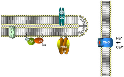
The principal components of the vertebrate phototransduction cascade, rhodopsin, transducin (Gαβγ), and cGMP phosphodiesterase-6 (PDE6), reside on the disc membranes within a specialized outer segment compartment of rod photoreceptor cells.
Step 2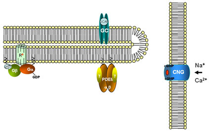
Photoexcited rhodopsin (R*) binds transducin and stimulates exchange of GTP for GDP on the Gα-subunit.
Step 3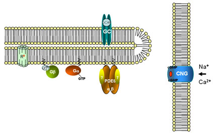
GαGTP dissociates from Gβγ and R*.
Step 4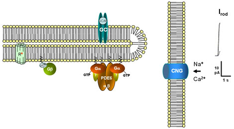
GαGTP activates PDE6 by displacing the inhibitory γ-subunits (Pγ) from the PDE catalytic core (PDE6αβ). cGMP hydrolysis by activated PDE6 results in closure of cGMP-gated (CNGC) channels in the plasma membrane
Step 5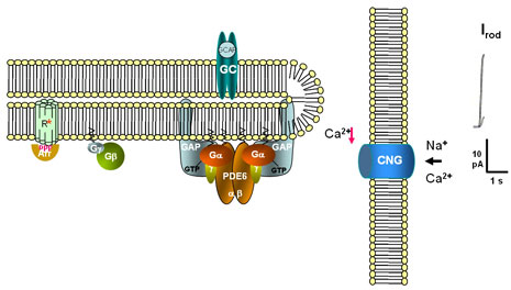
The turn-off phase of the visual signal is determined by reactions controlling the lifetimes of photoexcited rhodopsin (R*) and activated transducin. The ability of R* to continue to activate transducin is blocked by phosphorylation of the C-terminal Ser and Thr residues by rhodopsin-kinase and binding of arrestin to phosphorylated R*
The lifetime of GαGTP depends upon its intrinsic GTPase activity. Hydrolysis of GTP switches the Gα molecule to the inactive GDP-bound conformation. A GTPase-activating protein complex (GAP) comprising of a photoreceptor-specific member of the RGS (regulators of G protein signaling) family, RGS9-1, Gβ5L, and R9AP (RGS-9-1-anchor protein), accelerates inactivation of Gα. The Pγ-subunits potentiate the GAP-action of RGS9 by increasing the affinity between Gα and RGS9-1/Gβ5L/R9AP.
Step 6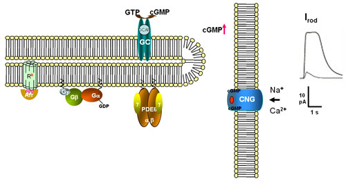
GαGDP re-associates with Gβγ and PDE6αβ is re-inhibited by Pγ. A decrease in the intracellular Ca2+ concentration resulting from the closure of cGMP-gated channels leads to activation of guanylate cyclase (GC) by specific Ca2+-binding GC-activating proteins (GCAPs) and restoration of cGMP levels. cGMP-gated channels reopen